Healthcare Professionals
Maraciclatide is for investigational use only and is not approved by the FDA or UK and European regulatory authorities.
⁹⁹ᵐTc-maraciclatide
Bringing Molecular Imaging to Inflammatory Arthritis
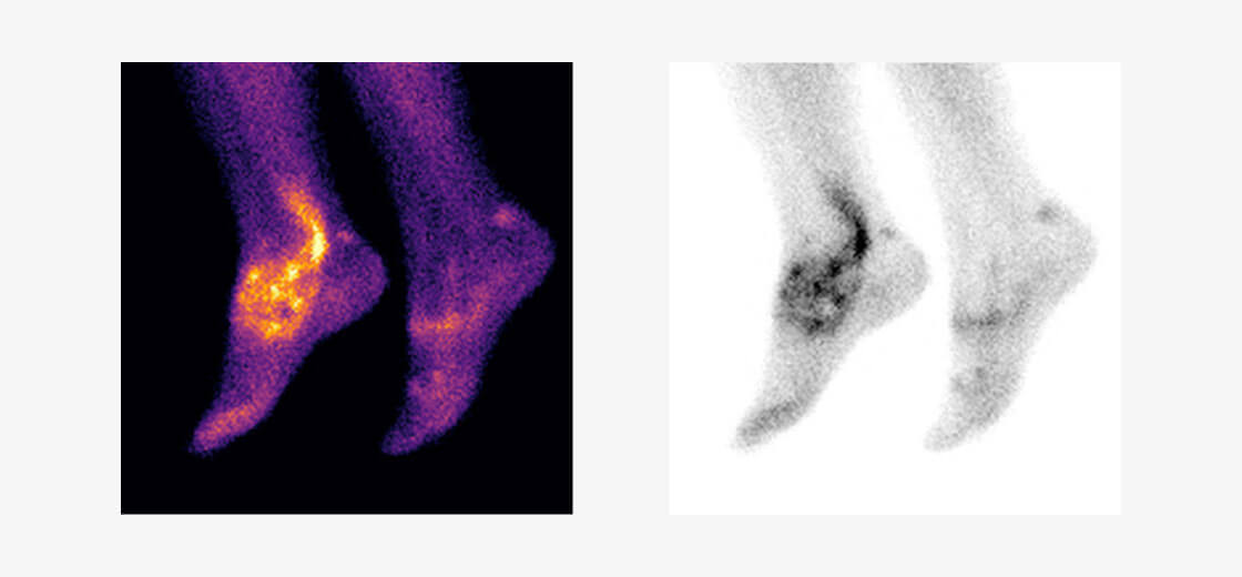
99mTc-maraciclatide is a radio-labelled tracer containing the RGD tripeptide motif (Arg-Gly-Asp) which binds with high affinity to αvβ3 integrin1, a cell-adhesion molecule which is up-regulated on activated vascular endothelial cells, activated macrophages and osteoclasts2. 99mTc-maraciclatide uptake in the joints has been shown to be highly correlated with Power Doppler Ultrasound (PDUS) in an initial proof of concept study in 5 patients with rheumatoid arthritis3 and subsequently in a study involving 50 patients4,5.
99mTc-maraciclatide planar imaging has the capacity to image the whole body, highlighting total synovial inflammatory load in a 20 minute acquisition without requiring the rheumatologist’s time.
99mTc-maraciclatide images are easy to interpret to the untrained observer and we are exploring how images might inform a patient’s view of their condition and treatment.
References
1 Ebenhan T, Kleynhans J, Zeevaart JR, et al. Non-oncological applications of RGD-based single-photon emission tomography and positron emission tomography agents. Eur J Nucl Med Mol Imaging 2020; https://doi.org/10.1007/s00259-020-04975-9
2 Eliceiri BP, Cheresh DA. The role of alphav integrins during angiogenesis: insights into potential mechanisms of action and clinical development. J Clin Invest 1999;103(9):1227-30.
3 Attipoe L, Chaabo K, Wajed J, et al. Imaging neoangiogenesis in rheumatoid arthritis (INIRA): whole-body synovial uptake of a 99mTc-labelled RGD peptide is highly correlated with power Doppler ultrasound. Ann Rheum Dis 2020;79:1254-1255.
4 Attipoe L, Garrood T, Subesinghe S, et al. Imaging Neoangiogenesis in Rheumatoid Arthritis (INIRA): Whole-Body Synovial Uptake of a 99mTc-Labelled RGD Peptide Is Highly Correlated with Power Doppler Ultrasound [abstract]. Arthritis Rheumatol 2019;71(suppl 10).
5 Attipoe L, Subesinghe S, Blanco-Gil C, et al. Imaging neoangiogenesis in rheumatoid arthritis (INIRA): whole-body synovial uptake of a 99mTc-labelled RGD peptide is highly correlated with power Doppler ultrasound. Ann Rheum Dis 2020;79, supplement 1:110.
Seracam (hybrid gamma optical camera) is for investigational use only and has not been cleared or approved by the FDA or UK and European regulatory authorities.
Seracam
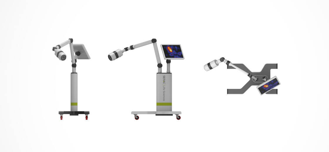
Point of care molecular imaging
- Small camera head can be placed closer to the patient than conventional large detectors which improves image resolution/acquisition times.
- Small footprint and fast acquisition to increase capacity in the Nuclear Medicine Department by providing an alternative for imaging small organs and structures without requiring an additional, dedicated camera room.
- Lightweight, portable design makes it easy for one person to move the camera around the Department, through hospital corridors, in the tight confines of the ICU, and around the patient’s bed.
- Imaging at the patient’s bedside (e.g. paediatric department, ICU) improves care and eliminates the need to bring the patient and nursing staff to the Nuclear Medicine Department.
Improving the Patient Experience
- Easy to position camera head making it possible to image patients in whatever position is most comfortable for them (e.g. sitting in a chair or wheelchair, or lying down on a bed or gurney).
- A less intimidating experience for the patient than with conventional SPECT cameras, making it ideal for claustrophobic and paediatric patients.
- Automatically co-registered gamma and optical images facilitates added information on uptake in relation to surface anatomy improving communication on investigational findings with the patient.
Read more
Bugby SL, et al. Investigation of an SFOV hybrid gamma camera for thyroid imaging. Physica Medica 2016;32:290–296.